Home » Without Label » Human Bone Anatomy Chart / Chicken Skeleton Labeled | Skeleton labeled, Skeleton ... / An excellent addition to anatomical models in the classroom or doctors office.available in 3 versions,
Human Bone Anatomy Chart / Chicken Skeleton Labeled | Skeleton labeled, Skeleton ... / An excellent addition to anatomical models in the classroom or doctors office.available in 3 versions,
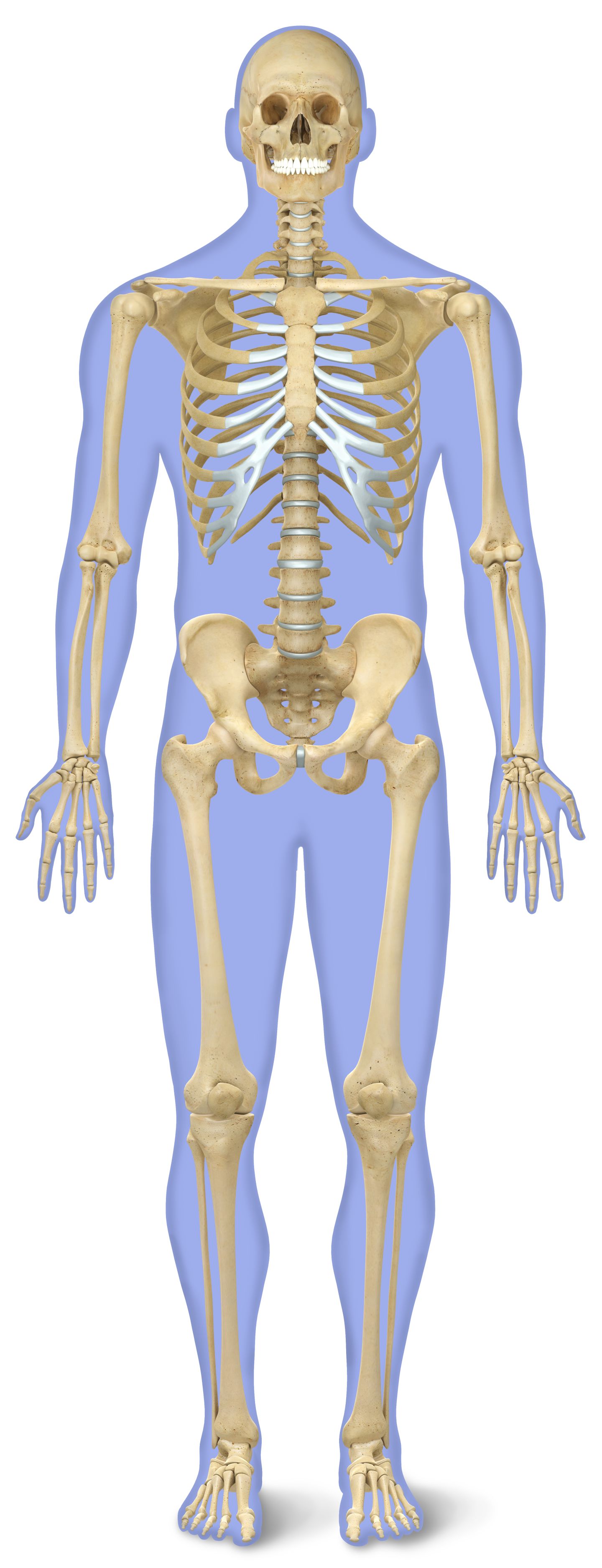
Human Bone Anatomy Chart / Chicken Skeleton Labeled | Skeleton labeled, Skeleton ... / An excellent addition to anatomical models in the classroom or doctors office.available in 3 versions,. The bones of the axial skeleton act as a hard shell to protect the internal organs—such as the brain and the heart—from damage caused by external forces. It is composed of 300 bones at birth, but later decreases to 80 bones in the axial skeleton and 126 bones in the appendicular skeleton. Human body (206) axial skeleton (80) appendicular skeleton (126) skull (28) torso (52) upper extremity (32 x 2 = 64) lower extremity (31 x 2 = 62) paired bones (11 x 2 = 22) nasal lacrimal inferior nasal concha maxiallary zygomatic temporal palatine parietal malleus incus stapes paired bones (12 x 2 = 24) rib 1 rib 2 rib 3 rib 4 rib 5 rib 6 Anatomy chart of human bones for medicine design. The femur is a type of long bone located in the thigh and is the largest bone of the skeletal system.
And most of the nasal cavity is composed of parts of the ethmoid bone. Zygote body is a free online 3d anatomy atlas. The bones of the appendicular skeleton provide support and flexibility at the joints and anchor the muscles that move the limbs. The femur and/or hip may fracture secondary to trauma, so understanding the femur bone anatomy is important. They make an impact and customers reviews have been great.
Human Male Anatomy 3D Model MAX OBJ FBX C4D LWO LW LWS MA ... from img-new.cgtrader.com Zygote body is a free online 3d anatomy atlas. Vector isolated flat illustration of skull and bones in body. Human anatomy diagrams show internal organs, cells, systems, conditions, symptoms and sickness information and/or tips for healthy living. Explore the anatomy systems of the human body! This diagram depicts printable human skeleton 744×1022 with parts and labels. Anatomy chart of human bones for medicine and health care themes design. Human skeleton labeled human skeleton bones human skeleton anatomy human body anatomy human anatomy and physiology anatomy of the body skeleton figure skeleton model human skull. Check out pictures and diagram related to bones, organs, senses, muscles and much more.
It is composed of 300 bones at birth, but later decreases to 80 bones in the axial skeleton and 126 bones in the appendicular skeleton.
Vector isolated flat illustration of skull and bones in body. Skeleton bone diagram of hip, foot. Human skeleton labeled human skeleton bones human skeleton anatomy human body anatomy human anatomy and physiology anatomy of the body skeleton figure skeleton model human skull. Anatomical wall charts and posters from 3b scientific® are ideal for teaching human anatomy, patient education and medical studies! Halloween, medical, educational or science banner human anatomy chart stock illustrations Posted on may 28, 2014 by admin. See more ideas about human anatomy, muscle anatomy, anatomy. Anatomy chart of human bones for medicine design. This diagram depicts human skeleton with parts and labels. The human skeleton bone and bone growth bone is living tissue, and, as such, can grow and remodel during a person's lifetime. This diagram depicts printable human skeleton 744×1022 with parts and labels. Bone zygomatic bone maxilla mandible nasal bones perpendicular plate of ethmoid nasal conchae note the nasal bones only make up a small portion of the bridge of the nose, most of the external nose is cartilage. It is composed of 300 bones at birth, but later decreases to 80 bones in the axial skeleton and 126 bones in the appendicular skeleton.
Bone diagram forehead (frontal bone) nose bones (nasals) cheek bone (zygoma) upper jaw (maxilla) lower jaw (mandible) breast bone (sternum) upper arm bone (humerus) lower arm bone (ulna) thigh bone (femur) collar bone (clavicle) toe bones (phalanges) ankle bones (tarsals) kneecap (patella) shin bone The anatomy of the femur can be divided into proximal, central, distal, and posterior parts. View, isolate, and learn human anatomy structures with zygote body. Human body (206) axial skeleton (80) appendicular skeleton (126) skull (28) torso (52) upper extremity (32 x 2 = 64) lower extremity (31 x 2 = 62) paired bones (11 x 2 = 22) nasal lacrimal inferior nasal concha maxiallary zygomatic temporal palatine parietal malleus incus stapes paired bones (12 x 2 = 24) rib 1 rib 2 rib 3 rib 4 rib 5 rib 6 This diagram depicts human bone diagram.
science anatomy scan of human body organs and bones Motion ... from d2v9y0dukr6mq2.cloudfront.net Altogether, the skeleton makes up about 20 percent of a person's body weight. They make an impact and customers reviews have been great. Human skeleton labeled human skeleton bones human skeleton anatomy human body anatomy human anatomy and physiology anatomy of the body skeleton figure skeleton model human skull. Related posts of human back bone chart muscles and bones in the arm. Welcome to innerbody.com, a free educational resource for learning about human anatomy and physiology. The anatomy of the femur can be divided into proximal, central, distal, and posterior parts. Check out pictures and diagram related to bones, organs, senses, muscles and much more. And most of the nasal cavity is composed of parts of the ethmoid bone.
The three types of bone cells are the osteoblasts, which are responsible for bone growth;
This diagram depicts printable human skeleton 744×1022 with parts and labels. Posted in diagrams | tagged all bones, human skeleton, skelet, skeleton human eye featured. Learn more about human anatomy with these free resources. The anatomy of the femur can be divided into proximal, central, distal, and posterior parts. This entry was posted in anatomy and tagged bone, bone anatomy, bone chart, bone charts, bone diagram, bone diagrams, bone graph, bone graphic, bone graphs, bone image, bone infographic, bone plot, bone table, bones anatomy, bones diagram, human bone, human bones, human bones diagram by admin. This diagram depicts human skeleton with parts and labels. See more ideas about human anatomy, muscle anatomy, anatomy. The bones of the appendicular skeleton provide support and flexibility at the joints and anchor the muscles that move the limbs. Bone diagram forehead (frontal bone) nose bones (nasals) cheek bone (zygoma) upper jaw (maxilla) lower jaw (mandible) breast bone (sternum) upper arm bone (humerus) lower arm bone (ulna) thigh bone (femur) collar bone (clavicle) toe bones (phalanges) ankle bones (tarsals) kneecap (patella) shin bone The femur and/or hip may fracture secondary to trauma, so understanding the femur bone anatomy is important. It is composed of 300 bones at birth, but later decreases to 80 bones in the axial skeleton and 126 bones in the appendicular skeleton. Related posts of human back bone chart muscles and bones in the arm. Human body (206) axial skeleton (80) appendicular skeleton (126) skull (28) torso (52) upper extremity (32 x 2 = 64) lower extremity (31 x 2 = 62) paired bones (11 x 2 = 22) nasal lacrimal inferior nasal concha maxiallary zygomatic temporal palatine parietal malleus incus stapes paired bones (12 x 2 = 24) rib 1 rib 2 rib 3 rib 4 rib 5 rib 6
Human anatomy diagrams show internal organs, cells, systems, conditions, symptoms and sickness information and/or tips for healthy living. Posted in diagrams | tagged all bones, human skeleton, skelet, skeleton human eye featured. Altogether, the skeleton makes up about 20 percent of a person's body weight. Explore the anatomy systems of the human body! This diagram depicts human skeleton with parts and labels.
Number of Bones in Human Body | Skeleton Facts | DK Find Out from res.cloudinary.com Halloween, medical, educational or science banner human anatomy chart stock illustrations Human skeleton labeled human skeleton bones human skeleton anatomy human body anatomy human anatomy and physiology anatomy of the body skeleton figure skeleton model human skull. Welcome to innerbody.com, a free educational resource for learning about human anatomy and physiology. This diagram depicts printable human skeleton 744×1022 with parts and labels. This diagram depicts human bone diagram. Anatomical wall charts and posters from 3b scientific® are ideal for teaching human anatomy, patient education and medical studies! The human skeletal system consists of all of the bones, cartilage, tendons, and ligaments in the body. View, isolate, and learn human anatomy structures with zygote body.
Human anatomy diagrams show internal organs, cells, systems, conditions, symptoms and sickness information and/or tips for healthy living.
The bones of the axial skeleton act as a hard shell to protect the internal organs—such as the brain and the heart—from damage caused by external forces. The osteoclasts, which are active in bone resorption; This diagram depicts human bone diagram. This diagram depicts printable human skeleton 744×1022 with parts and labels. Bones human skull diagram, chart of human bones back view, human bone chart pdf, human bones chart, human bones diagram with names, bone, bones human skull diagram. The anatomy of the femur can be divided into proximal, central, distal, and posterior parts. View, isolate, and learn human anatomy structures with zygote body. Human body (206) axial skeleton (80) appendicular skeleton (126) skull (28) torso (52) upper extremity (32 x 2 = 64) lower extremity (31 x 2 = 62) paired bones (11 x 2 = 22) nasal lacrimal inferior nasal concha maxiallary zygomatic temporal palatine parietal malleus incus stapes paired bones (12 x 2 = 24) rib 1 rib 2 rib 3 rib 4 rib 5 rib 6 This entry was posted in anatomy and tagged bone, bone anatomy, bone chart, bone charts, bone diagram, bone diagrams, bone graph, bone graphic, bone graphs, bone image, bone infographic, bone plot, bone table, bones anatomy, bones diagram, human bone, human bones, human bones diagram by admin. Posted on may 28, 2014 by admin. The bones of the appendicular skeleton provide support and flexibility at the joints and anchor the muscles that move the limbs. Muscles and bones in the arm 12 photos of the muscles and bones in the arm muscles and bones. Check out pictures and diagram related to bones, organs, senses, muscles and much more.